Overview Of Anatomy Of The Ear
The human ear, organ of hearing that detects and analyzes sound by transduction (or the conversion of sound waves into electrochemical impulses) and maintains the sensation of balance. It is the organ of the senses that allows us to listen. Hearing can be defined as the perception of sound energy through the brain and the central nervous system.
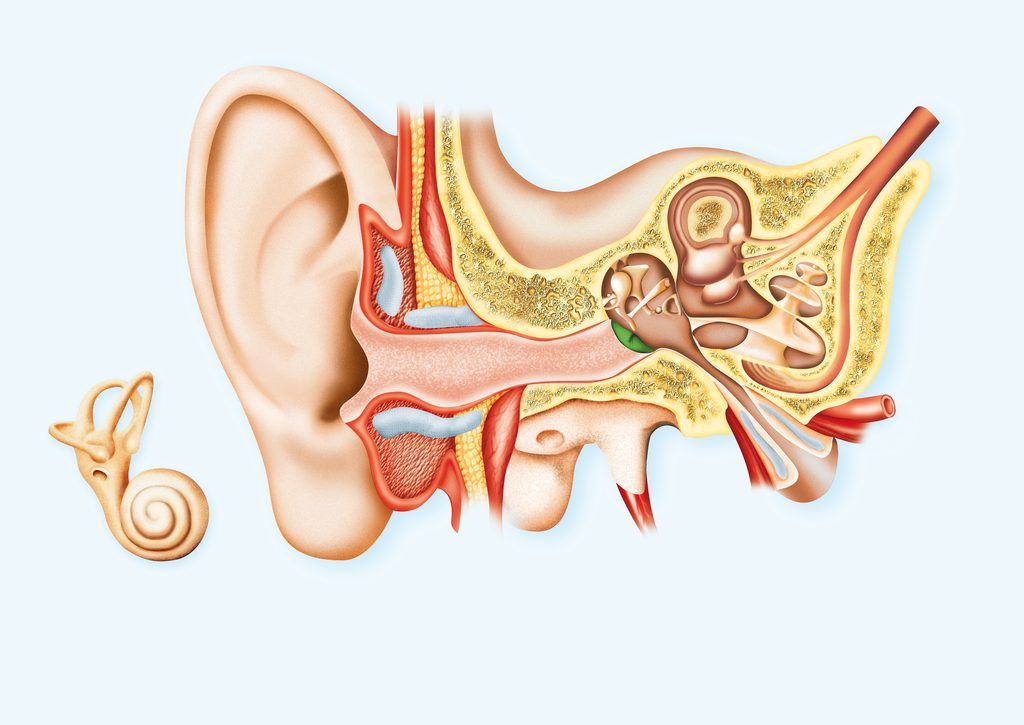
External ear
This part acts as a funnel to drive the vibrations of the air to the eardrum. It also has the function of sound localization. The external ear consists of the auricular pavilion and the auditory canal.
Pinna
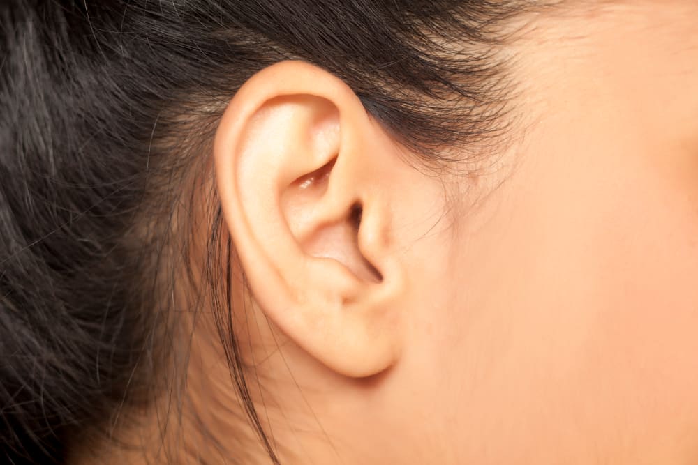
The auricular pavilion is a prominent flap covered by the skin located on the side of the head and is the visible part of the ear externally. It is formed and supported by the cartilage with the exception of the earlobe. It picks up sound waves and channels them to the external auditory canal through patterns formed in the ear pavilion known as whorls and voids.
Its shape also partially protects the sound waves that come close to the ear from the back, allowing a person to know if a sound comes directly from the front or not.
Auditory canal
The auditory canal is approximately 3 cm long in adults and slightly S-shaped. It is supported by the cartilage in its opening, and by bone for the rest of its length. The skin covers the canal and contains glands that produce secretions that mix with dead skin cells to produce cerumen.
The cerumen, together with the fine hairs that protect the entrance to the auditory canal, helps to prevent particles in the air from reaching the internal parts of the auditory canal. Where the eardrum may accumulate or injure and interfere with hearing. The cerumen usually dries out and falls out of the canal. However, sometimes it can become compact and interrupt hearing.
Middle ear
It is located between the outer and inner ear. It is separated from the ear canal of the outer ear by the tympanic membrane (the eardrum). It works to transfer the vibrations of the eardrum to the fluid of the inner ear. This transfer of sound vibrations is possible through a chain of small mobile bones, called ossicles, that extend through the middle ear and their corresponding small muscles.
Tympanic membrane (eardrum)
It is commonly known as the eardrum and separates the ear canal from the middle ear. It is approximately 1 cm in diameter and is slightly concave on its outer surface. It vibrates freely in response to sound. The membrane is very innervated, so it is very sensitive to pain. For the membrane to move freely when the air hits it, the pressure of the resting air on both sides of the tympanic membrane should be the same.
The outside of the membrane is exposed to atmospheric pressure through the auditory tube, so that the cavity in which it is located, called tympanic cavity, is continuous with the cells in the jaw.
Normally, the auditory tube is elongated and closed, but swallowing, yawning and chewing, opens, allowing air to enter or exit the tympanic cavity. This opening of the auditory tube allows the air pressure in the middle ear to equilibrate with atmospheric pressure so that the pressures on both sides of the tympanic membrane equal each other.
Auditory ossicles and muscles
The tympanic cavity contains the bones and the smallest muscles of the body. The bones are also called auditory ossicles and connect the eardrum to the inner ear. From the outermost to the innermost, the bones are called hammer, anvil, and stirrup.
The hammer is attached to the eardrum. It has a handle that adheres to the inner surface of the eardrum and a head that is suspended from the wall of the tympanic cavity. The anvil is connected to the hammer on the side closest to the eardrum, and to the abutment on the side closest to the inner ear.
The stirrup has a bow and a platform. This footplate is held by a piece of annular tissue in an opening called the oval window, which is the entrance to the inner ear. The stapedium is the muscle of the inner ear that is inserted into the stapes. The tympanic tensor is the muscle of the inner ear that is inserted into the hammer. With which, it converts the vibrations of the sound wave into the fluid movement of the inner ear.
Inner ear
It is the deepest part of the entire ear and is located in a place known as the bony labyrinth. This is a labyrinth bordered by a network of fleshy tubes known as the membranous labyrinth.
A fluid cushion, called perilymph, is found between the bony labyrinth and the membranous labyrinth, while a fluid called endolymph is found within the membranous labyrinth itself. Inside the inner ear, there is a chamber called the vestibule, which plays an important role in the sense of balance.
Cochlea
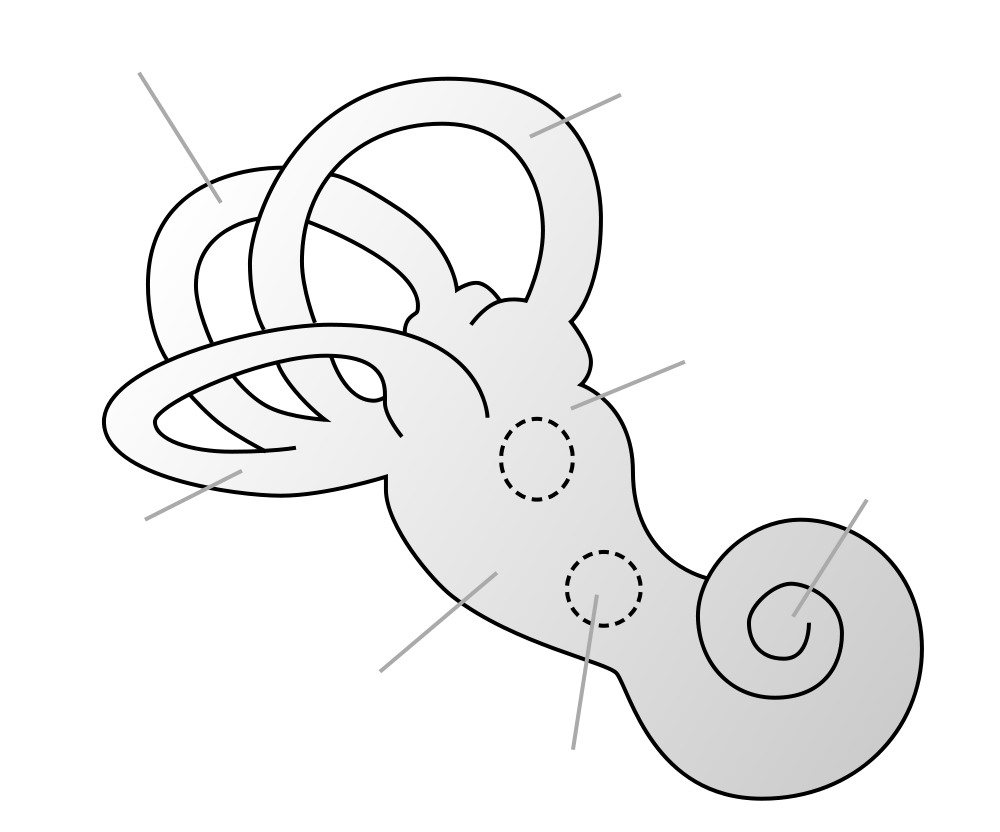
From the hall comes the cochlea, which is sometimes referred to as the organ of hearing. It is the part of the complete ear that really converts the vibrations of sound into auditory perception. The cochlea is shaped like a spiral in the shape of a snail. It is about 9 mm wide at the base and 5 mm high, and it is wrapped around a section of cancellous bone called modiolo.
The modiolo has the shape of a screw whose threads form a spiral platform that supports the cochlea, which is fleshy and unable to sustain itself.
Cochlea Chambers
The cochlea contains three chambers filled with liquid separated by membranes. The upper chamber, the scala hall and the lower chamber, the scala tympani, are full of perilinfa. The scala tympani is covered by a secondary tympanic membrane. The intermediate chamber is the middle scala or the cochlea duct. It is full of endolymph, instead of perilymph.
Organ of Corti
This organ is supported by a membrane called the basilar membrane. It is about the size of a pea, and acts as a transducer, converting the vibration into nerve impulses. It has hair cells and support cells. The hair cells have long, rigid microvilli called stereocilia on their apical surfaces.
The microvilli are fine structures like hairs in the cells that help increase the area of the cell surface. In addition to these stereocilia, it has a gelatinous membrane called the tectorial membrane. Four rows of hair cells rotate along the organ of Corti. These outer hair cells adjust the response of the cochlea to different sound frequencies to allow the inner hair cells to function more accurately.